Services
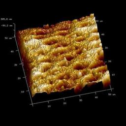
Atomic Force Microscope (AFM) facilitates the imaging and analysis of biological surfaces with little or no sample preparation. AFM operates in a near field with a sharp probe by scanning, enabling characterization of three-dimensional surface morphology with resolution at a nano-metric scale. Atomic Force Microscope (AFM) aids to analyze the topography of the biological hard tissue surfaces and may quantitatively determine the surface roughness.
NanoDMA analysis: Dentin or bone may deform according to a combination of the viscoelastic properties and exhibit time-dependent strain. The complex modulus is a measure of the resistance of a material to dynamic deformation. The nanoDMA analysis is able to assess the dampening (or viscous) behavior of the tissue at a nano-metric scale, as mechanical properties are very sensitive to the tissue structural and histological changes.
Raman spectroscopy is a nondestructive technique which may allow the analysis of the biochemical nature, molecular structure and emission spectroscopies of tested tissues. Through the Raman spectrometer/microscope, individual spectral bands arising from the mineral and organic constituents of dentin/bone may be identified. Micro-Raman single point spectra (figure 1) or mapping techniques appeared to offer a powerful method for a direct analysis of the phosphate peak heights at the dentin surface (figure 2) or at the bonded resin polymer-dentin interface (figure 3). Evolution of these analyses may be used to assess for tissue remineralization. Various methods of multivariate analysis, like hierarchical cluster analysis (HCA), principal component analysis (PCA), Classical Least Squares (CLS) Fitting or Clustering K-Means (KMC) have been established for evaluating two dimensional data (figure 4).
By integrating RAMAN, AFM and nano-DMA morpho-chemio and nanomechanical properties analysis can be obtained, related and mapped.


